|
|
Small Bowel Lipoma
General Considerations
- Small bowel tumors are relatively rare, accounting for only 2% of all GI malignancies
- Most lipomas of the GI tract occur in the colon, the small bowel being the 2nd most common site and accounting for about 25%
- Usually solitary but may be multiple
- Most small bowel lipomas are found in the ileum
- Most often submucosal and may be pedunculated
Clinical Findings
- If small, usually no symptoms
- When symptoms occur, they are non-specific and may include
- Abdominal pain
- Constipation
- Diarrhea
- Nausea and vomiting
- GI hemorrhage (may be occult), especially when over 2 cm
- Obstruction (in up to 30% of symptomatic patients)
- Lead point of intussusception
Imaging Findings
- Conventional radiographs will be normal except when there is obstruction of the bowel
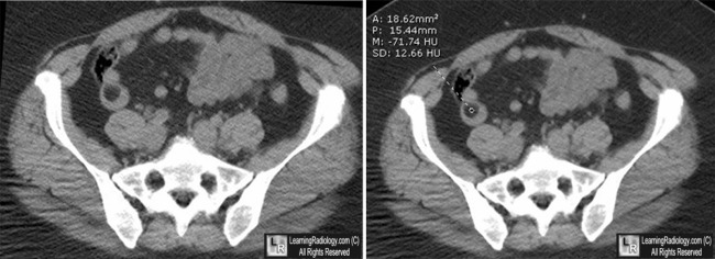
- CT will show a low density mass in the wall of the small bowel
- Homogeneous mass with Hounsfield units between −80 and –120
- Upper GI series may show a submucosal mass with obtuse borders that may have barium collections in central ulcerations
- The Lipoma may be noted to change its shape on different views owing to its pliability
Differential Diagnosis
- Adenomas
- Carcinoid tumor
- Intestinal polyposis syndromes
Treatment
- If symptomatic, they may be surgically removed

Lipoma of Small Bowel. The white arrow points to sharply marginated filling defect in the distal small bowel. The mass measures -71 Hounsfield units, consistent with fat density.
For these same photos without the arrows, click here
For more information, click on the link if you see this icon 
Imaging and Findings of Lipomas of the Gastrointestinal Tract. WM Thompson .AJR:184, April 2005
|
|
|
